Tuesday, April 26, 2016

TPA NETWORK SERVICE HOSPITALS IN HYDERABAD INFORMATION


 TPA BEST SERVICE HOSPITALS IN HYDERABAD INFORMATION 


  • MEDI ASSIST INDIA PVT. LTD. 
  • DEDICATED HEALTH SERVICES.
  • E-MEDITEK HEALTH SOLUTIONS PVT. LTD.
  • MD INDIA PVT. LTD.
  • UNITED HEALTH CARE SERVICES PVT.LTD.
  • VIDAL ( TTK )HEALTH CARE SERVICES PVT. LTD.
  • HERITAGE HEALTH CARE SERVICES.
  • VIPUL MED INDIA PVT. LTD.
  • FAMILY HEALTH PLAN LTD.
  • GENESIS INDIA PVT. LTD.
  • HEALTH CARE  PVT. LTD.
  • ICICI LOMBARD PVT .LTD.
  • MAXBUPA PVT. LTD.
  • R CARE.
  • RELIGARE
  • UHC (UNITED HEALTH CARE).
  • BAJAJ ALLIANZ GENERAL INSURANCE.
  • FUTURE GENERAL INSURANCE.
  • UNIVARSAL SOMPO GENERAL INSURANCE.
  • PARAMOUNT HEALTH SERVICE.

  • CENTRAL GOVERNMENT  HEALTH SCHEME.
  • TELANGANA STATE GOVERNMENT  HEALTH SCHEME.
  • ANDHRAPRADESH STATE GOVERNMENT HEALTH SCHEME.
  • APCPDCL
  • TSCPDCL
  • TSRTC 
  • APSRTC
  • ESI CORPORATION.
  • Call 9989704849

Monday, April 25, 2016

Aarogyasri Free Surgery ACL Tear in Hyderabad- Telangana


 SURGERY FOR  ACL TEAR IN HYDERABAD PRIVATE HOSPITALS UNDER  AAROGYASRI 


మన తెలంగాణ/ఆంద్ర్హప్రదేశ్ రాష్ట్ర ప్రభుత్వాలు బి.పి.యల్. కుటుంబాలకు మాత్రమే కాకుండా పింక్ కార్డు

 దారులు కూడా ఆరోగ్యశ్రీ  ఉచిత ఆరోగ్యసేవలను అందించడంకోసం ఏర్పాటు చేసిందే తెలంగాణ

 ఆరోగ్యశ్రీ...... ఇప్పటికే  ఈ పధకం ద్వార అత్యవసర సమయంలో కొన్ని కోట్ల కుటుంబాలను

 రోడ్డున పడకుంట ఆదుకుంటున్న ఏకైక గొప్ప ప్రభుత్వ పథకం ఆరోగ్యశ్రీ ...

 తెలంగాణ/ ఆంద్ర్హప్రదేశ్  రాష్ట్రంలోని ఏ ఓక్క కుటుంబం కూడా ఆపద సమయంలో ఆర్దికంగా 

నష్టపోకుండా ఉండాలనే దృడసంకల్పంతో ఈ ఆరోగ్యశ్రీ పధకంను రాష్ట్ర ప్రభుత్వం అమలు చేస్తుంది

 ఒక్క మాటలో చెప్పాలంటే పేదోడికి ప్రవేట్ వైద్యశాలలు

రెడ్ కార్పెట్ వేసి మరి ఆరోగ్య సేవలను అందిస్తున్నారు అంటే అది కేవలం ఆరోగ్యశ్రీ పధకం ద్వారానే
.
 దురదృష్టకరమైన విషయం ఏమిటంటే ఇప్పటికి ఆరోగ్యశ్రీ సేవలను ఎలాపొందాలో తెలియక పోవడం.


ప్రవేట్ హాస్పిటల్లలో  ఆరోగ్యశ్రీ కార్డు, వైట్ రేషను కార్డు లేకపోయీనా ఆరోగ్యశ్రీ సేవలను పొందవచ్చు.. 9989704849

                               This surgery CASHLESS for all  resindent in Telangana and                                            Andhra Pradesh only under  AROGYASRI services

         REQUIRED DOCUMENTS;
  RATION CARD  /  VOTER CARD / AADAR CARD / DRIVING LICENSE / SCHOOL ID CARD / AAROGYASREE CARD /  SC,ST CAST CERTIFICATE . ETC..
          .ANY ONE DOCUMENT REQUIRED.

IN HYDERABAD PRIVATE AAROGYASRI HOSPITALS FREE SURGERY CALL.

ACL ( Anterior Cruciate Ligament )


Ligament  a band of fibrous tissue, structure connecting bones, which strength joints cruciate its of knee “x” like structure.

They are four ligaments in the knee joint.          
1. Lateral collateral ligament,     2. Medial collateral ligament,
 3, Anterior cruciate ligament,   4, Posterior cruciate ligament.

 Anterior cruciate ligament  This ligament  is most commonly damaged, often in association with the tears of medial or lateral collateral ligaments .  Commonly, it occurs as a result of twisting force on a semi – flexed knee.

 With increasing sporting activities, injuries to the knee ligaments are on the increase.
 Knee ligaments are injured most often from indirect, twisting or bending forces on the knee.

 The ACL can be injured in several ways……


  •   Changing direction rapidly.
  •   Stopping suddenly.
  •   Slowing down while running.
  •   Landing from a jump incorrectly and football tackles.


Symptoms:


  •   Loss of full range of motion.
  •   Pain with swelling knee.
  •   Tenderness along the knee joint.
  •   If go walking discomport.

ACL Tear once brake will not heal without surgery.


           Knee Replacement

Staff nurse vacancy in NIMS Hyderabad TELANGANA

NIMS Staff Nurse Recruitment 2016 Latest Nursing Jobs in Telangana Nizam’s Institute of Medical Sciences Hyderabad Telangana Are Inviting Applications for Junior Staff Nurse Internees,From Eligible and Qualified Candidates to work Contract Basis,candidates who are looking,Waiting for Staff Nurse Jobs NIMS Staff Nurse Recruitment 2016 Latest Nursing Jobs in Telangana Eligible and qualified candidates i.e, B.Sc Nursing,and Should be registered or General Nursing Midwifery Jobs details,Jobs Description,Number of Vacancies,Age limit,Salary for Month, Details Eligibility qualification,Educational qualifications,Desirable qualification,Mode of apply Time and Date of the registration Information about NIMS Staff Nurse Recruitment 2016 Latest Nursing Jobs in Telangana related details given below:
I.Details of the Posts:
Sr. NoName of PostVacancyPay Scale
1Staff Nurse100 (One Hundred )
Rs.17000/-
II. Eligibility Criteria:  B.Sc Nursing/GNM
III.Age Limit:
  • Minimum Age:- 18 years
  • Maximum Age:- 34 years(5 Year Relaxation for ST/SC)
IV. Application Fee:
  • General/OBC candidates: 1000/-
  • SC/ST/PHD candidates: 500/-
V.How to Apply: Steps to apply-
  • Firstly, eligible candidates are required to visit online the official website of www.nims.edu.in
  • Now, Click on the “’Apply”link.
  • Fill the form and deposit the application fee as mentioned above in the form of DD or Cash.
  • Submit to Below Address
VI.Address:
   Executive Registrar,
       Nizam’s institute of Medical Sciences,
      Panjagutta, Hyderabad, Telangana

VII.Mode of Selection:
  • Direct Recruitment
  • Interview
VII. Important Date:
  • Starting Date for Online Registration:- 13-04-2016
  • Last Date for Online Registration:- 30-04-2016

Friday, April 22, 2016

aarogyasri services use for pink card holders also


పింక్ కార్డు దారులు కుడా ఆరోగ్యశ్రీ సేవలను

పొందవచ్చు

అత్యవసర సమయంలో మన శరీరంలోని 

చేతులు, కాళ్ళు, మోకాలి చిప్ప

వెన్నుపూస, విరిగినప్పుడు మరియు 

మెదడు, గుండె శాస్త్ర చికిస్థలను ప్రైవేటు హాస్పిటల్లో ఉచిత ఆరోగ్యశ్రీ సేవలను

పొందుట గురించి


  మన తెలంగాణ/ఆంధ్రప్రదేశ్  రాష్ట్ర ప్రభుత్వాలు బి.పి.యల్. కుటుంబాలకు 

మాత్రమే కాకుండా పింక్ కార్డు దారులు కూడా ఆరోగ్యశ్రీ  ఉచిత 

ఆరోగ్యసేవలను అందించడంకోసం ఏర్పాటు చేసిందే తెలంగాణ/ఆంధ్రప్రదేశ్ 

ఆరోగ్యశ్రీ......ఇప్పటికే  ఈ పధకం ద్వార అత్యవసర సమయంలో కొన్ని కోట్ల

 కుటుంబాలను రోడ్డున పడకుంట ఆదుకుంటున్న ఏకైక గొప్ప ప్రభుత్వ

 పథకం ఆరోగ్యశ్రీ ...


 తెలంగాణ/ ఆంధ్రప్రదేశ్   రాష్ట్రంలోని ఏ ఓక్క కుటుంబం కూడా ఆపద 

సమయంలో ఆర్దికంగా నష్టపోకుండా ఉండాలనే దృడసంకల్పంతో ఈ

ఆరోగ్యశ్రీ పధకంను రాష్ట్ర ప్రభుత్వం అమలు చేస్తుంది


 ఒక్క మాటలో చెప్పాలంటే పేదోడికి ప్రవేట్ వైద్యశాలలు

రెడ్ కార్పెట్ వేసి మరి ఆరోగ్య సేవలను అందిస్తున్నారు అంటే అది కేవలం

 ఆరోగ్యశ్రీ పధకం ద్వారానే
.
 దురదృష్టకరమైన విషయం ఏమిటంటే ఇప్పటికి ఆరోగ్యశ్రీ సేవలను 

ఎలాపొందాలో తెలియక పోవడం.

ప్రవేట్ హాస్పిటల్లలో  ఆరోగ్యశ్రీ కార్డు, వైట్ రేషను కార్డు లేకపోయీనా ఆరోగ్యశ్రీ

 సేవలను పొందవచ్చు..

        ఎలాగంటే.?....... ఏదైనా ప్రమాదవషత్తు కాలో,చెయ్యో విరిగి ప్రైవేటు 

హాస్పిటల్ లో చేరినప్పుడు మిమ్ముల్ని పరీక్ష చేసిన డాక్టర్ విరిగిన బాగానికి 

శాస్త్ర చికిస్థ చేయాల్సి వస్తే ఆ శాస్త్ర చికిస్థ ఆరోగ్యశ్రీ జాబితాలో ఉంటే  మీ వద్ద 

 ఆరోగ్యశ్రీ కార్డు, వైట్ కార్డు లేకపోయిన ఏ దేని ప్రభుత్వ సంస్థల  గుర్తింపు 

కార్డు అంటే ఉదాహరణకు...

 పింక్ కార్డు / పాన్  కార్డు  / ఆధార్ కార్డు / వోటర్  కార్డు  / డ్రైవింగ్ లైసెన్సు / 

బ్యాంకుపాస్ బుక్ / పోస్టాఫీసు పాస్ బుక్  /  పాస్ పోర్ట్ / స్కూల్ ఐ డి కార్డు / 

వికలాంగుల ద్రువికరణ పత్రము / కార్మిక శాఖా గుర్తింపు కార్డు.. / చిన్న

 పిల్లలకు బర్త్ సర్టిఫికేట్ మొ.. వి. 

వీటిలో ఏదేని ఒక దృవపత్రం మరియు డాక్టర్ దృవీకరణ పత్రాలను తీసుకోని

  రోగి నేరుగా  సి యం క్యాంపు ఆఫీస్ కి వెళ్లి  సి యం సి ఓ రిఫరల్ కార్డు 

పొందవచును....... ఈ సి యం సి ఓ కార్డు ను ప్రైవేటు హాస్పటల్ లోని 

ఆరోగ్యశ్రీ కౌంటర్ లో ఇస్తే సరిపోతుంది... పూర్తిగా  వైద్య సేవలను  ఉచితంగా 

పొందవచ్చును...... 9989704849

Thursday, April 14, 2016

best service aarogyasri hospitals in hyderabad information

FREE SURGERY FOR FRACTURE OF 

CLOSED PATELLA

HYDERABAD 

PRIVATE AAROGYASRI HOSPITALS 


INFOMATION;

మన తెలంగాణ/ఆంద్రప్రదేశ్ రాష్ట్ర ప్రభుత్వాలు బి.పి.యల్. కుటుంబాలకు మాత్రమే కాకుండా పింక్ కార్డు

 దారులు కూడా ఆరోగ్యశ్రీ  ఉచిత ఆరోగ్యసేవలను అందించడంకోసం ఏర్పాటు చేసిందే తెలంగాణ/

 ఆంద్రప్రదేశ్  ఆరోగ్యశ్రీ...... ఇప్పటికే  ఈ పధకం ద్వార అత్యవసర సమయంలో కొన్ని కోట్ల

కుటుంబాలను రోడ్డున పడకుంట ఆదుకుంటున్న ఏకైక గొప్ప ప్రభుత్వ పథకం ఆరోగ్యశ్రీ ...

 తెలంగాణ/ ఆంద్రప్రదేశ్   రాష్ట్రంలోని ఏ ఓక్క కుటుంబం కూడా ఆపద సమయంలో ఆర్దికంగా 

నష్టపోకుండా ఉండాలనే దృడసంకల్పంతో ఈ ఆరోగ్యశ్రీ పధకంను రాష్ట్ర ప్రభుత్వం అమలు చేస్తుంది

 ఒక్క మాటలో చెప్పాలంటే పేదోడికి ప్రవేట్ వైద్యశాలలు

రెడ్ కార్పెట్ వేసి మరి ఆరోగ్య సేవలను అందిస్తున్నారు అంటే అది కేవలం ఆరోగ్యశ్రీ పధకం ద్వారానే
.
 దురదృష్టకరమైన విషయం ఏమిటంటే ఇప్పటికి ఆరోగ్యశ్రీ సేవలను ఎలాపొందాలో తెలియక పోవడం.


ప్రవేట్ హాస్పిటల్లలో  ఆరోగ్యశ్రీ కార్డు, వైట్ రేషను కార్డు లేకపోయీనా ఆరోగ్యశ్రీ సేవలను పొందవచ్చు..


                               THIS SURGERY FREE FOR ALL TELANGANA AND                                               ANDRHAPRADHESH   RESIDENCE.....ONLY... UNDER  AROGYASRI

         REQUIRED DOCUMENTS;
  RATION CARD  /  VOTER CARD / AADAR CARD / DRIVING LICENSE / SCHOOL ID CARD / AAROGYASREE CARD / CAST CERTIFICATE .
          .ANY ONE DOCUMENT REQUIRED.


I

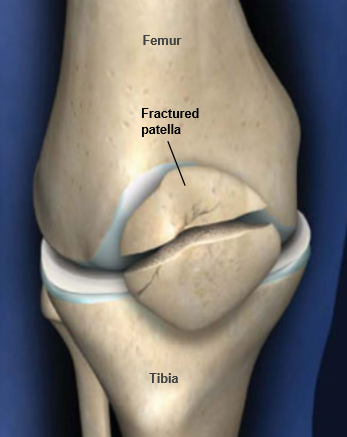
Patellar (Kneecap) Fractures
Because your kneecap (patella) acts like a shield for your knee joint, it can easily be broken. Falling directly onto your knee, for example, is a common cause of patellar fractures.
These fractures are serious injuries and often require surgery to heal. Over the long term, they may cause arthritis in the knee.
Anatomy
The patella is a small bone located in front of your knee joint — where the thighbone (femur) and shinbone (tibia) meet. It protects your knee and connects the muscles in the front of your thigh to your tibia.
The ends of the femur and the undersides of the patella are covered with a slippery substance called articular cartilage. This helps the bones glide smoothly along each other as you move your leg
Statistics
legtellar fractures account for about 1% of all fractures. They are most common in people who are 20 to 50 years old. Men are twice as likely as women to fracture the kneecap.
Types of Patellar Fractures
Patellar fractures vary. The kneecap can crack just slightly, or can be broken into many pieces.
A break in the kneecap can happen at the top, center, or lower part of the bone. Sometimes, fractures occur in more than one area of the kneecap.
This x-ray of the knee taken from the side shows a patella that has been fractured in three places.
Stable fracture. This type of fracture is nondisplaced. The broken ends of the bones meet up correctly and are aligned. In a stable fracture, the bones usually stay in place during healing.
A vertical, stable fracture.
This x-ray of the knee taken from the front shows the faint line of a vertical, stable fracture of the patella.
Displaced fracture. When a bone breaks and is displaced, the broken ends are separated and do not line up. This type of fracture often requires surgery to put the pieces back together.
A transverse fracture with some displacement.
This x-ray of the knee taken from the side shows a significant gap (displacement) between the broken pieces of the patella.
Comminuted fracture. This type of break is very unstable. The bone shatters into three or more pieces.
A comminuted fracture of the patella.
Open fracture. In this type of fracture, the skin has been broken and exposes the bone. These injuries often involve much more damage to the surrounding muscles, tendons, and ligaments. Open fractures have a higher risk for complications and take a longer time to heal.
Cause
Patellar fractures are most commonly caused by a direct blow, such as from a fall or motor vehicle collision. The patella can also be fractured indirectly. For example, your thigh muscles can contract so violently that it pulls the patella apart.

Symptoms
The major symptoms of a patellar fracture include pain and swelling in the front of the knee. Additional symptoms include:
  • Bruising
  • Inability to straighten the knee
  • Inability to walk

Doctor Examination
After discussing your symptoms and medical history, your doctor will examine your knee. The edges of the fracture can often be felt through the skin, particularly if the fracture is displaced. Your doctor will also check for hemarthrosis. This is swelling deep inside the joint that is usually a result of bleeding caused by the fracture.

Tests
X-rays are the most common and widely available diagnostic imaging technique. They create images of dense structures, like bone, so are particularly useful in showing fractures. X-rays are important for showing front and side views of the fracture.
Although rare, a person may be born with extra bones in the patella that have not grown together. This is called bipartite patella and may be mistaken for a fracture. X-rays help to identify bipartite patella. Many people have bipartite patella in both knees, so your doctor may take an x-ray of your other knee, as well.

Treatment

Nonsurgical Treatment

If the pieces of broken bone have not been displaced by the force of the injury, you may not need surgery. Casts or splints may be used to keep your knee straight. This will keep the broken ends in proper position while they heal.
You will not be able to put any weight on your leg until the bone is completely healed. This may take 6 to 8 weeks, and perhaps longer. Most people use crutches during this period.

Surgical Treatment

If the patella has been pulled apart (displaced), you will most likely need surgery. Fractured patellar bones that are not close together often have difficulty healing or may not heal. The thigh muscles that attach to the top of the patella are very strong and can pull the broken pieces out of place during healing.
Timing of surgery. If the skin around your fracture has not been broken, your doctor may recommend waiting until any abrasions have healed before having surgery. Open fractures, however, expose the fracture site to the environment. They urgently need to be cleansed and require immediate surgery.
Procedure. The type of procedure performed often depends on the type of fracture you have. Before the surgery, your doctor will discuss your procedure with you, as well as any potential complications.
A figure-of-eight tension band holds the transverse fracture together.
  • Transverse fracture.These two-part fractures are most often fixed in place using pins and wires and a "figure-of-eight" configuration tension band. The figure-of-eight band presses the two pieces together.
  • This procedure is best for treating fractures that are located near the center of the patella. Fracture pieces at the ends of the kneecap are too small for this procedure. Breaks that are in many pieces can be overcompressed by the tension band.
    Another approach to a transverse fracture is to secure the bones using small screws, wires, and pins. In many cases, these wires and pins will need to be removed about a year or two after the surgery.
  • Comminuted fracture. In some cases, either the top or the bottom of the patella can be broken into several small pieces. This type of fracture happens when the kneecap is pulled apart from the injury, then crushed from falling on it.
  • Because the bone fragments are too small to be fixed back into place, they will be removed. Your doctor will attach the loose tendon to the remaining patellar bone.
    If the kneecap is broken in many pieces at its center (and they are separated), your doctor may use a combination of wires and screws to fix it. Removing small portions of the kneecap may also have good results. Complete removal of the kneecap is a last resort in treating a comminuted fracture.

PLEASE CONTACT FOR MORE INFORMATION .... 

24x7 SEVICE

 GRM SEVA SAMITHI.....

G. MALLESH .. 9989704849.

Wednesday, April 13, 2016

ehs best service knee replacement hospitals in hyderabad information

  EMPLOY HEALTH SCHEME  

 BEST SERVICE KNEE REPLACEMENT HOSPITALS IN HYDERABAD


---------------------------------------------------------------------------------

Saturday, April 9, 2016

Free surgery for fracture in ( shine bone)closed Tibia Fibula in Hyderabad private hospitals information


Free surgery for fracture in closed tibia  fibula in hyderabad private AAROGYASRI hospitals information

మన తెలంగాణ/ఆంద్ర్హప్రదేశ్ రాష్ట్ర ప్రభుత్వాలు బి.పి.యల్. కుటుంబాలకు మాత్రమే కాకుండా పింక్ కార్డు దారులు కూడా ఆరోగ్యశ్రీ  ఉచిత ఆరోగ్యసేవలను అందించడంకోసం ఏర్పాటు చేసిందే తెలంగాణ ఆరోగ్యశ్రీ...... ఇప్పటికే  ఈ పధకం ద్వార అత్యవసర సమయంలో కొన్ని కోట్ల కుటుంబాలను రోడ్డున పడకుంట ఆదుకుంటున్న ఏకైక గొప్ప ప్రభుత్వ పథకం ఆరోగ్యశ్రీ ...

 తెలంగాణ/ ఆంద్ర్హప్రదేశ్  రాష్ట్రంలోని ఏ ఓక్క కుటుంబం కూడా ఆపద సమయంలో ఆర్దికంగా నష్టపోకుండా ఉండాలనే దృడసంకల్పంతో ఈ ఆరోగ్యశ్రీ పధకంను రాష్ట్ర ప్రభుత్వం అమలు చేస్తుంది

 ఒక్క మాటలో చెప్పాలంటే పేదోడికి ప్రవేట్ వైద్యశాలలు
రెడ్ కార్పెట్ వేసి మరి ఆరోగ్య సేవలను అందిస్తున్నారు అంటే అది కేవలం ఆరోగ్యశ్రీ పధకం ద్వారానే
.
 దురదృష్టకరమైన విషయం ఏమిటంటే ఇప్పటికి ఆరోగ్యశ్రీ సేవలను ఎలాపొందాలో తెలియక పోవడం.


ప్రవేట్ హాస్పిటల్లలో  ఆరోగ్యశ్రీ కార్డు, వైట్ రేషను కార్డు లేకపోయీనా ఆరోగ్యశ్రీ సేవలను పొందవచ్చు..

                               This surgery free for all  resindent in Telangana and                                            Andhra Pradesh only under  AROGYASRI services

         REQUIRED DOCUMENTS;
  RATION CARD  /  VOTER CARD / AADAR CARD / DRIVING LICENSE / SCHOOL ID CARD / AAROGYASREE CARD / CAST CERTIFICATE .
          .ANY ONE DOCUMENT REQUIRED.

IN HYDERABAD CORPORATE HOSPITALS FREE SURGERY CALL.
PLEASE CONTACT FOR MORE INFORMATION .... GRM SEVA SAMITHI .....
G. MALLESH ..9989704849.

Tibia (Shinbone) Shaft Fractures
The tibia, or shinbone, is the most common fractured long bone in your body. The long bones include the femur, humerus, tibia, and fibula. A tibial shaft fracture occurs along the length of the bone, below the knee and above the ankle.
Because it typically takes a major force to break a long bone, other injuries often occur with these types of fractures.
Anatomy
The lower leg is made up of two bones: the tibia and fibula. The tibia is the larger of the two bones. It supports most of your weight and is an important part of both the knee joint and ankle joint.
The tibia is the larger bone in your lower leg. Tibial shaft fractures occur along the length of the bone.

Types of Tibial Shaft Fractures
The tibia can break in several ways. The severity of the fracture usually depends on the amount of force that caused the break. The fibula is often broken as well.
Common types of tibial fractures include:
Stable fracture: This type of fracture is barely out of place. The broken ends of the bones basically line up correctly and are aligned. In a stable fracture, the bones usually stay in place during healing.
Displaced fracture: When a bone breaks and is displaced, the broken ends are separated and do not line up. These types of fractures often require surgery to put the pieces back together.
Transverse fracture: This type of fracture has a horizontal fracture line. This fracture can be unstable, especially if the fibula is also broken.
Oblique fracture: This type of fracture has an angled pattern and is typically unstable. If an oblique fracture is initially stable or minimally displaced, over time it can become more out of place. This is especially true if the fibula is not broken.
Spiral fracture: This type of fracture is caused by a twisting force. The result is a spiral-shaped fracture line about the bone, like a staircase. Spiral fractures can be displaced or stable, depending on how much force causes the fracture.
Comminuted fracture: This type of fracture is very unstable. The bone shatters into three or more pieces.
Open fracture: When broken bones break through the skin, they are called open or compound fractures. For example, when a pedestrian is struck by the bumper of a moving car, the broken tibia may protrude through a tear in the skin and other soft tissues.
Open fractures often involve much more damage to the surrounding muscles, tendons, and ligaments. They have a higher risk for complications and take a longer time to heal.
Closed fracture: With this injury, the broken bones do not break the skin. Although the skin is not broken, internal soft tissues can still be badly damaged. In extreme cases, excessive swelling may cut off blood supply and lead to muscle death, and in rare cases, amputation.

Cause
High-energy collisions, such as an automobile or motorcycle crash, are common causes of tibial shaft fractures. In cases like these, the bone can be broken into several pieces (comminuted fracture).
Sports injuries, such as a fall while skiing or running into another player during soccer, are lower-energy injuries that can cause tibial shaft fractures. These fractures are typically caused by a twisting force and result in an oblique or spiral type of fracture.

Symptoms
The most common symptoms of a tibial shaft fracture are:
  • Pain
  • Inability to walk or bear weight on the leg
  • Deformity or instability of the leg
  • Bone "tenting" the skin or protruding through a break in the skin
  • Occasional loss of feeling in the foot
Doctor Examination
It is important that your doctor knows the circumstances of your injury. For example, if you fell from a tree, how far did you fall? It is just as important for your doctor to know if you sustained any other injuries and if you have any other medical problems, such as diabetes. Your doctor also needs to know if you take any medications.
After discussing your symptoms and medical history, your doctor will do a careful examination. He or she will assess your overall condition, and then focus on your leg. Your doctor will look for:
  • Obvious deformity such as angulation or shortening (the legs are not the same length)
  • Breaks in the skin
  • Contusions (bruises)
  • Swelling
  • Bony prominences under the skin
  • Instability (some patients may retain a degree of stability if the fibula remains intact or the fracture is incomplete)
After the visual inspection, your doctor will feel along your leg to see if there are abnormalities of the tibia. If you are awake and alert, your doctor will test your sensation and muscle strength by asking you to move your toes and see if you can feel different areas over your foot and ankle.

Tests
Other tests that may help your doctor confirm your fracture include:

X-rays

X-rays can show if the bone is broken and whether there is displacement (the gap between broken bones). They can also show how many pieces of bone there are. X-rays are also useful for identifying the involvement of the knee or ankle joint, and the presence of a fibula fracture.

Computed tomography (CT) scan

After reviewing your x-rays, your doctor may recommend a CT scan of your leg. This is often done if there is a question of the fracture extending into either the knee or ankle joint. A CT scan shows a cross-sectional image of your limb. It can provide your doctor with valuable information about the severity of the fracture.

Treatment
In planning your treatment, your doctor will consider several things, including:
  • The cause of your injury
  • Your overall health
  • The severity of your injury
  • The extent of soft tissue damage

Nonsurgical Treatment

Nonsurgical treatment may be recommended for patients who:
  • Are poor surgical candidates due to their overall health problems
  • Are less active, so are better able to tolerate small degrees of angulation or differences in leg length
  • Have closed fractures with only two major bone fragments and little displacement (gap)
Initial treatment. Most injuries cause some swelling for the first few weeks. Your doctor may initially apply a splint to provide comfort and support. Unlike a full cast, a splint can be tightened or loosened, and allows swelling to occur safely. Once the swelling goes down, your doctor will consider a range of treatment options.
Cast and functional brace. One proven nonsurgical treatment method is to immobilize the fracture in a cast for initial healing. After weeks in the cast, it can be replaced with a functional brace made of plastic and fasteners. The brace will provide protection and support until healing is complete. The brace allows you to take it off for hygiene issues and for physical therapy.

Surgical Treatment

Your doctor may recommend surgery for your fracture if it is:
  • An open fracture with wounds that need monitoring
  • Extremely unstable because of many bone fragments and large degrees of displacement
  • Not healed with nonsurgical methods
Intramedullary nailing. The current most popular form of surgical treatment for tibial fractures is intramedullary nailing. During this procedure, a specially designed metal rod is inserted from the front of the knee down into the marrow canal of the tibia. The rod passes across the fracture to keep it in position.
Intramedullary nailing.
Intramedullary nails come in various lengths and diameters to fit most tibia bones. The intramedullary nail is screwed to the bone at both ends. This keeps the nail and the bone in proper position during healing.
Intramedullary nailing allows for strong, stable, full-length fixation. The technique also makes it more likely that the position of the bone obtained at the time of surgery will be maintained when compared with casting or external fixation.
Intramedullary nailing is not ideal for fractures in children and adolescents because care must be taken to avoid crossing the bone's growth plates.
Plates and screws. Tibial shaft fractures were once routinely treated with plate and screw constructs. These tools are reserved for fractures in which intramedullary nailing may not be possible or optimal, such as certain fractures that extend into either the knee or ankle joints.
During this type of procedure, the bone fragments are first repositioned (reduced) into their normal alignment. They are held together with special screws and metal plates attached to the outer surface of the bone.
External fixation. In this type of operation, metal pins or screws are placed into the bone above and below the fracture site. The pins and screws are attached to a bar outside the skin. This device is a stabilizing frame that holds the bones in the proper position so they can heal.
While external fixation has yielded some reasonable results, having implants outside the body has proven to be somewhat unpopular with many patients and physicians.

Image result for tibia fibula fracture

TPA NETWORK SERVICE HOSPITALS IN HYDERABAD INFORMATION

  TPA BEST SERVICE HOSPITALS IN HYDERABAD  INFORMATION  MEDI ASSIST INDIA PVT. LTD.  DEDICATED HEALTH SERVICES. E-MEDITEK HEA...